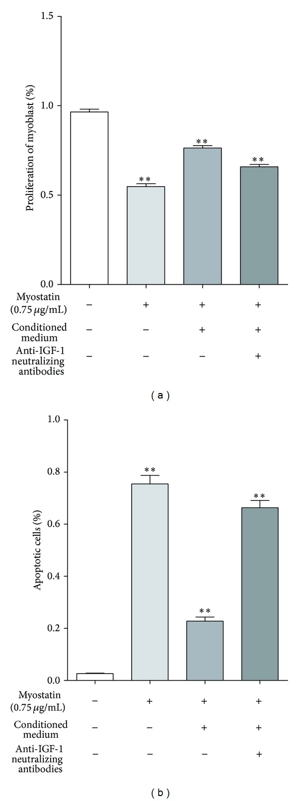
Figure 5.

(a) Effect of conditioned medium on myoblasts' proliferation is shown as percentage after 48 h myostatin exposure. Proliferation of murine myoblast cultured in 0.75 μg/mL myostatin significantly decreased to 54.8% (±3.2%, **P < 0.001), whereas conditioned medium of ASCs significantly increased myoblasts' proliferation under 0.75 μg/mL myostatin incubation to 76.4% (±2.7%, **P < 0.001). This protective effect was abolished after neutralization of IGF-1 ligand-receptor interaction and proliferation significantly decreased to 65.8% (±2.9%, **P < 0.001). (b) Myoblasts exposed to 0.75 μg/mL myostatin showed a significant increase in apoptosis rate to 75.5% (±8.7%) but were reduced to 22.8% (±4.2%) due to paracrine factors of ASCs conditioned medium (**P < 0.001). The protective effect of ASCs conditioned medium was abolished after blocking IGF-1 and its receptor whereas apoptotic rate increased to 66.3% (±7.3%) (**P < 0.001). Thus, IGF-1 secreted by ASCs accounts for 82.5% of the antiapoptotic effect of the CM (N = 3, n = 3).
