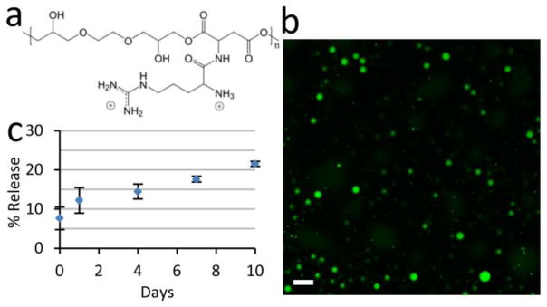
Figure 1. The coacervate controls the release of HB-EGF in a steady fashion.

(a) Chemical structure of the PEAD. The backbone of PEAD contains aspartic acid and ethylene glycol diglyceride, connected by biodegradable ester bonds. Arginine is conjugated to provide two cationic charges per repeating unit, giving PEAD strong affinity for polyanions. (b) Confocal fluorescent imaging of fluorescein-labeled coacervate shows spherical morphology with droplet diameters of 10–500 nm. Bar = 1 μm. (c) In vitro release profile of HB-EGF from the coacervate into saline over 10 days. Bars indicate means ± SD.
