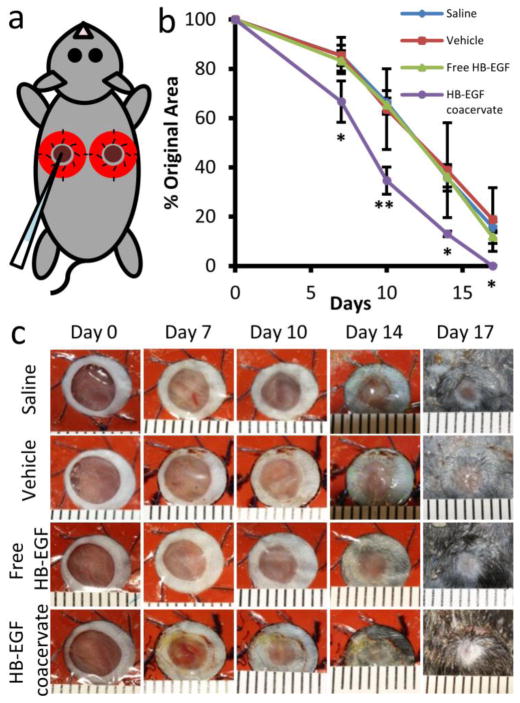
Figure 3. HB-EGF coacervate accelerates healing of full-thickness excisional wounds.
(a) Silicone donut-shaped splints, fixed with suture, surrounded the wounds and forced healing to occur by re-epithelialization. Wounds were treated immediately with 10 μl of group-specific solution by sterile pipet. (b) Wound closure over time, measured as percent of original area. Fourteen wounds were averaged for day 7 timepoint after which two animals were sacrificed per group for histology; the remaining 10 wounds per group were photographed until sacrifice on day 17. Bars indicate means ± SD. *P<0.05; **P<0.01. (c) Representative photographs of wounds treated with saline, vehicle (PEAD:heparin complex), 1 μg HB-EGF free or in the coacervate. Ruler units are mm.

