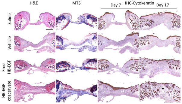
Figure 4. HB-EGF coacervate accelerates wound re-epithelialization and increases collagen content of granulation tissue.

Wound sections stained with hematoxylin and eosin (H&E) for general observation, Masson’s trichrome (MTS) for collagen, and immunohistochemically for cytokeratin (IHC-Cytokeratin) as a marker of epithelial cells. Representative light microscopy images of sections from the center of day 7 wounds are presented for all three staining methods, and of day 17 wound sections stained for IHC-Cytokeratin. Arrows indicate newly formed dermal appendages present within the granulation tissue. Bar = 500 μm.
