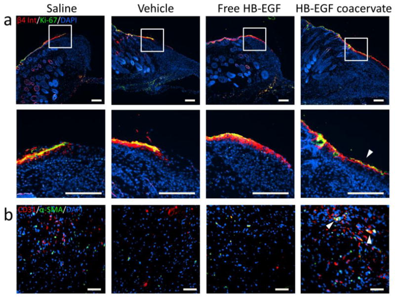
Figure 5. HB-EGF induces migration of epithelial cells with retained proliferative capability, and increases angiogenesis in wounds.

(a) Immunofluorescent staining of day 7 wound sections for β4 integrin (red) and Ki-67 (green) with DAPI (blue) nuclear staining. High magnification images of the wound margin reveal epithelial cells with retained proliferation potential (co-localization of markers; red + green = yellow). Arrow indicates significant migration of these dual-positive cells. Bars = 200 μm. (b) Immunofluorescent staining of day 17 wound sections for CD31 (red) and α-smooth muscle actin (green) with DAPI (blue) nuclear staining. Representative images of granulation tissue near the wound margin show endothelial cell and mural cell infiltration from the adjacent dermis. Arrows indicate co-localization of cells (red + green = yellow) as potential nascent vessels. Bars = 50 μm.
