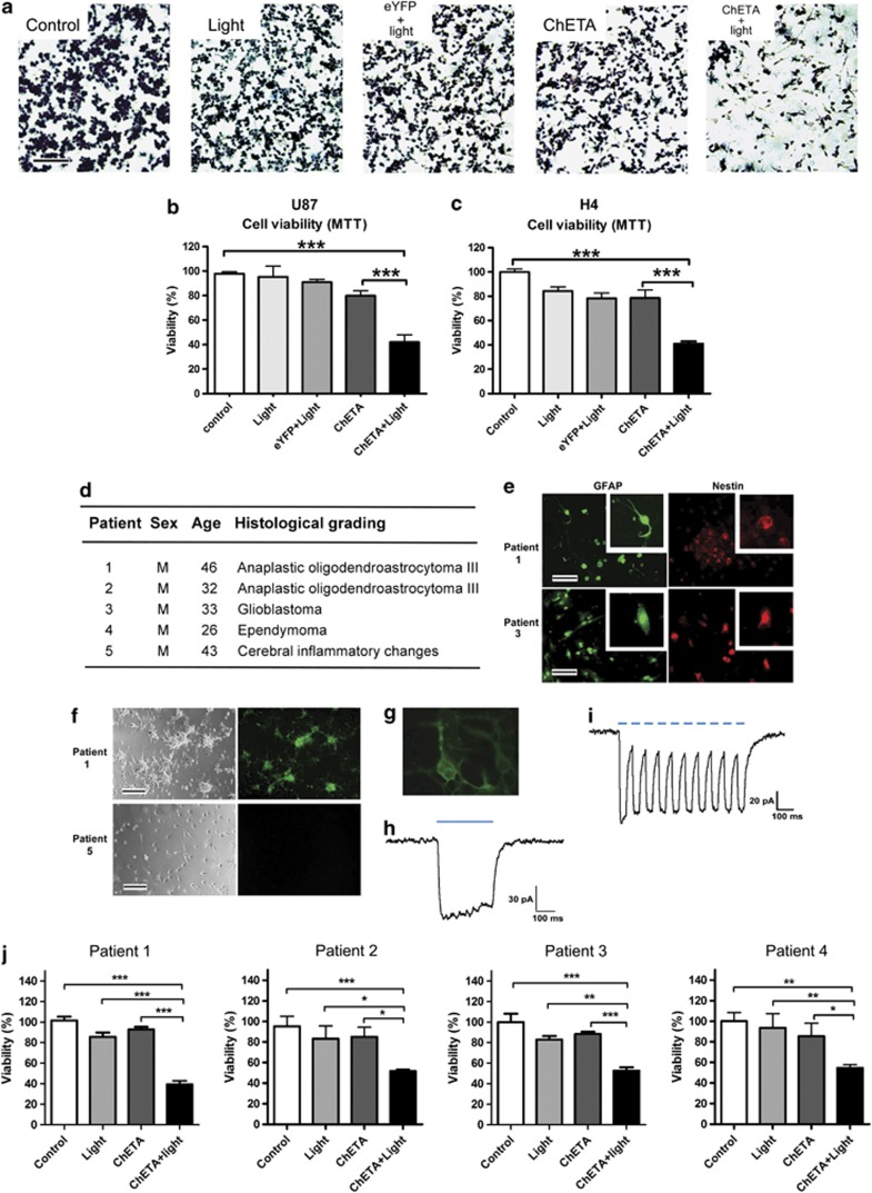
Figure 2.
ChETA gene expression and light stimulation reduced the viability of human glioma cells. (a) Crystal violet staining of the viable U87 cells 24 h after light stimulation; bar=100 μm. (b) Quantification of the viability of U87 cells in the indicated groups (n=15). (c) Quantification of the viability of H4 cells in the indicated groups (n=15). (d) Sex, age, and histological grading of the patients used for primary neural cell isolation and culture. (e) Immunofluorescence of GFAP and nestin on cultured primary human glioma cells (patients 1 and 3); bar=50 μm. (f) Representative ChETA expression in primary cultured human glioma cells (patient 1, right panel). No expression was observed in cells isolated from patient 5 who had no glioma; bars=50 μm. (g) A whole-cell patch clamp was used to record light-evoked photocurrents from representative ChETA-expressing primary human glioma cells (patient 1). (h) Light stimulation (500 ms) induced depolarization and the recovery current. (i) ChETA channel current spike trains induced by light pulses (50 ms, 10 Hz). (j) Quantification of primary human glioma cell viability in the control, light, ChETA, and ChETA+light groups isolated from patients 1–4, respectively. n=16. *P<0.05; **P<0.01; ***P<0.001

