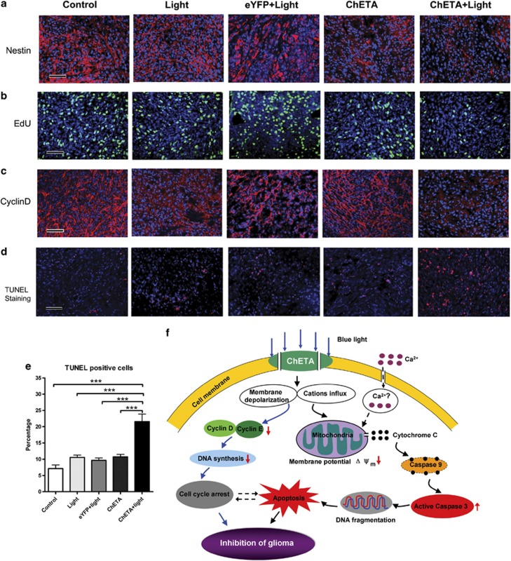
Figure 7.
Light stimulation inhibited the proliferation and induced the apoptosis of human glioma cells in vivo. (a) Immunostaining of nestin in the control, light, eYFP+light, ChETA, and ChETA+light groups. (b) EdU-positive cells (green) were observed in the control, light, eYFP+light, ChETA, and ChETA+light groups. Hoechst 33342-stained nuclei are shown in blue. (c) Immunostaining of cyclin D in the control, light, eYFP+light, ChETA, and ChETA+light groups. (d) TUNEL staining of apoptotic U87 cells in the control, light, eYFP+light, ChETA, and ChETA+light groups; Hoechst 33342-stained nuclei are shown in blue. (e) Quantification of the TUNEL-positive cells in the control, light, eYFP+light, ChETA, and ChETA+light groups. (f) Schematic drawing for the mechanism of the light-induced regulation of glioma cell proliferation and apoptosis. Bar=50 μm (a–d). ***P<0.001. The experiments were repeated for three times with similar results

