Figure 5.
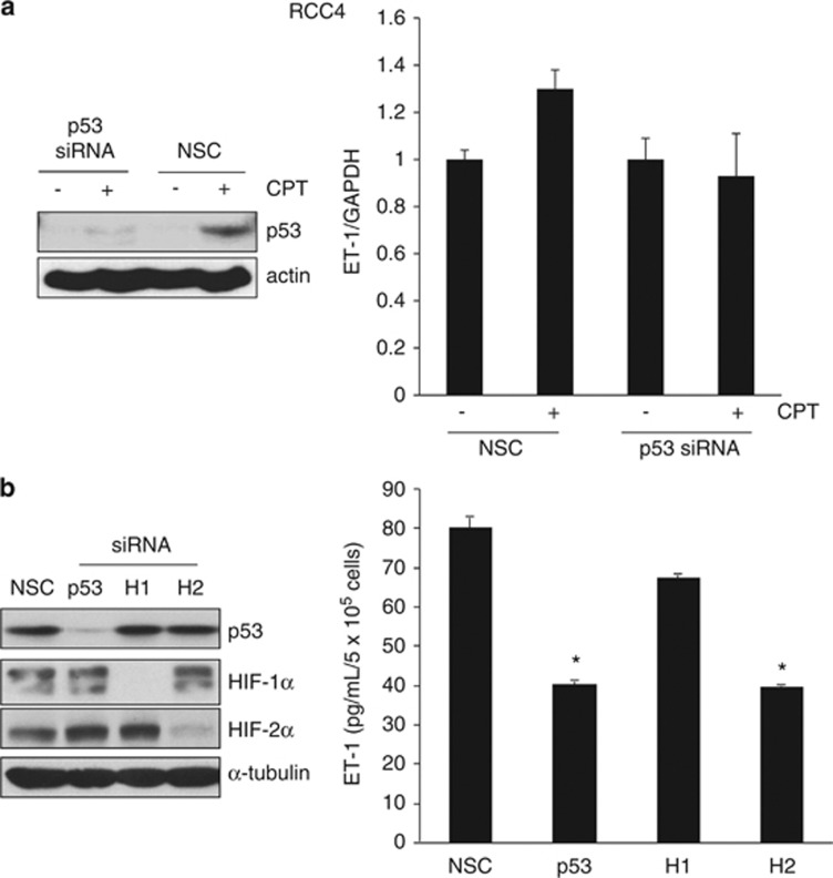
The p53-dependent regulation of ET-1 in RCC4 cells. (a) RCC4 cells were transfected with 10 nM siRNA to p53 or nonsilencing control (NSC) duplex for 24 h before addition of 2 μM CPT (+) or DMSO vehicle control (−) for a further 24 h. Panels, whole-cell lysates were assayed by western blot for p53 protein. Actin was used as a loading control. Graph, mRNA expression of ET-1 by real-time quantitative PCR relative to GAPDH. Mean±S.E. of duplicate values of one representative experiment is shown. (b) RCC4 cells were transfected with 10 nM siRNA to p53, HIF-1α (H1), or HIF-2α (H2) or NSC duplex for 24 h. Panels, whole-cell lysates were assayed by western blot for p53, HIF-1α and HIF-2α proteins. Tubulin was used as a loading control. Graph, conditioned media were harvested and secreted protein levels of ET-1 were determined by ELISA and normalized to cell number. Mean±S.E. of duplicate values of one representative experiment is shown. *P<0.05, t test compared with control

