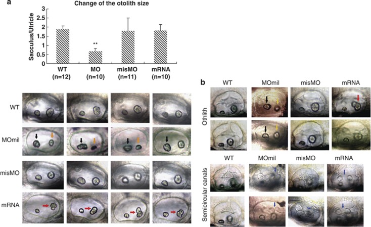
Figure 2.
Developmental defects of otoliths and semicircular canals caused by miles-apart dysregulation. Embryos were injected with 40 μM MOmil or 40 ng/ul mil-mRNA at the one-to four-cell stage and were observed at 2 d.p.f. (a) and 3 d.p.f. (b). Compared with WT embryos, abnormal otolith shapes emerged in the morphants at 2 d.p.f., such as enlarged and non-oval utricles (black arrows), and shrink sacculus (orange arrows) by MOmil injection (9/10). Although in misMO control group otoliths present shape (12/13) similar to that in WT (10/10). Embryos treated with miles-apart mRNA injection exhibit deformed, divided or syncytium-like sacculus (red arrows) and smaller utricles (black arrowhead) by miles-apart mRNA injection (11/12). A histogram indicates that variation of otolith sizes caused by miles-apart gene knockdown or overexpression using otolith area scanning, in which area ratio of sacculus to utricle was used as a sign. The means and S.D.'s are derived from the specified otic vesicles. **P-values <0.01. At 3 d.p.f. embryos (b) the top panel shows that the WT embryos developed normal semicircular canals, but deficient semicircular canals were appeared at the posterior part in the morphants (11/15), and deformation and missing occurred at the middle part of semicircular canals with mil-mRNA injection group (7/11). Few of the misMO group had the defect (3/13). Blue arrows show deficient semicircular canals. In addition, the enlarged non-oval utricles (black arrows) and shrink sacculus (orange arrows) by MOmil injection (11/15) and abnormal sacculuses by miles-apart mRNA injection (5/11, red arrows) were seen at 3 d.p.f. The images are left lateral view

