Figure 8.
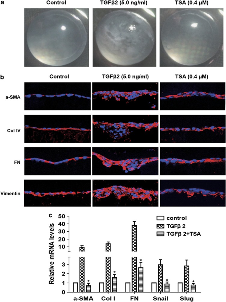
TSA abrogated TGFβ2-induced ASC in the whole lens culture semi-in vivo model. Lenses were cultured in the absence or presence of TGFβ2 with TSA (0.4 μM) or DMSO for 7 days. (a) The morphology of the lenses was photographed using a dissecting microscope. n=12. (b) The staining of frozen sections for α-SMA (red), Col IV (red), FN (red) and vimentin (red). Images were captured using a fluorescence confocal microscope. n=6. Bar, 20 μm. (c) The mRNA expression levels of α-SMA, Col I, FN, Snail and Slug in lenses were detected by real-time PCR. n=6. Gene levels were normalized to control glyceraldehyde 3-phosphate dehydrogenase. *P<0.05 versus TGFβ2 treated with DMSO group

