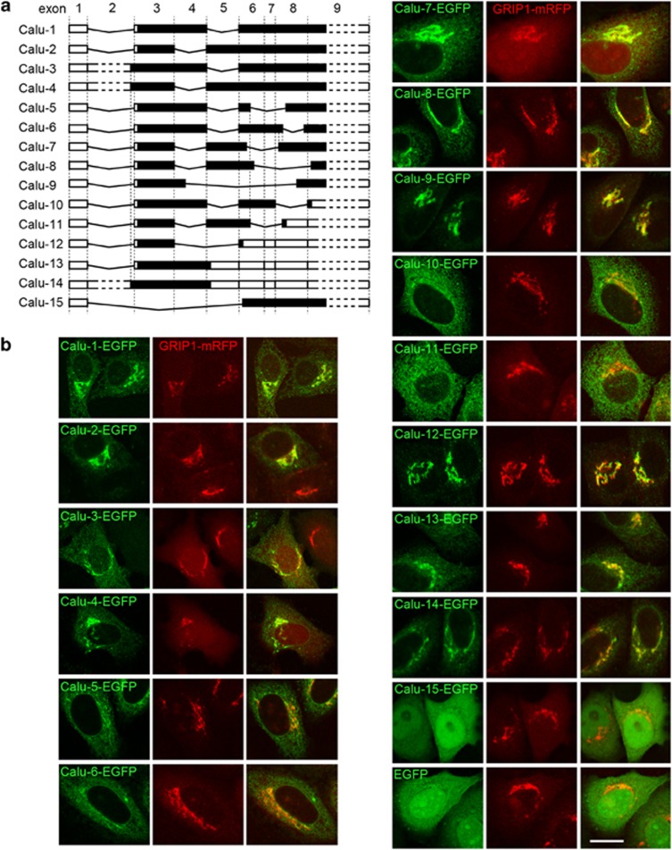
Figure 1.
Fifteen calumenin (Calu) isoforms and their subcellular localizations. (a) Schematic picture shows the exon organization of 15 Calu isoforms encoded by the CALU gene. The number at the top of the picture is the exon number. Black regions represent coding sequence, white regions represent untranslated mRNA sequence. (b) Subcellular localizations of the enhanced green fluorescent protein (EGFP) fusion proteins of Calu isoforms (green). GRIP1-mRFP (red) marks the Golgi apparatus. Bar, 10 μm

