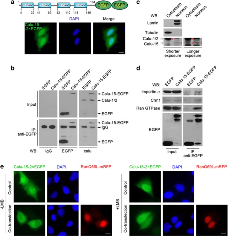
Figure 2.
Calumenin (Calu)-15 shuttles between the nucleus and the cytoplasm, mediated by importin-α, Ran GTPase, and Crm1. (a) Subcellular localization of Calu-15–2 × enhanced green fluorescent protein (EGFP) fusion protein (green). Nuclear DNA was stained with DAPI (4',6-diamidino-2-phenylindole; blue). Bar, 10 μm. The upper panel shows the domain architecture of Calu-15–2 × EGFP. (b) Immunoprecipitation (IP) with antibody against EGFP were performed in the lysates of HEK293T cells expressing Calu-15–EGFP, followed by immunoblotting (western blotting (WB)) with antibody against Calu-1/2 (calu). Antibody against EGFP (EGFP) as a positive control. IgG as a negative control. (c) Immunoblottings (WB) of endogenous Calu-15 in the cytoplasmic and nuclear fraction from HeLa cells. Asterisks indicate the band of Calu-1/2 or Calu-15. Tubulin and lamin, respectively, labels the cytoplasmic and nuclear fraction. (d) IP with antibody against EGFP was performed in the lysates of HEK293T cells expressing Calu-15–EGFP or EGFP, followed by immunoblotting (WB) with the indicated antibodies. (e) HeLa cells cotransfected with Calu-15–2 × EGFP and RanQ69L-mRFP were treated with (right panels) or without (left panels) leptomycin B (LMB) for 3 h, then fixed for observation. Nuclear DNA was stained with DAPI (blue). Bar, 10 μm

