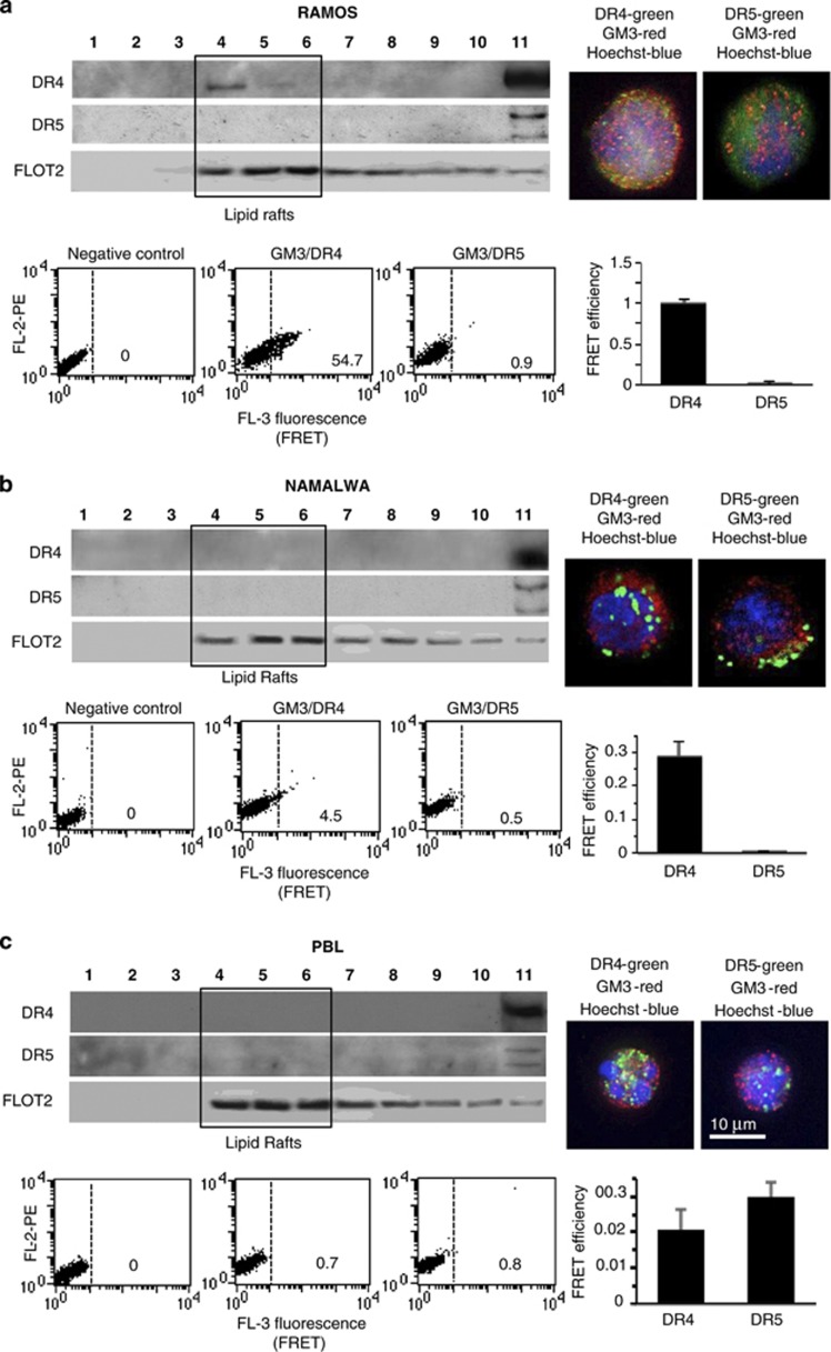
Figure 2.
Constitutive localization of TRAIL receptors into lipid rafts. Left panels. Western blot analysis of sucrose gradient fractions. Fractions obtained after sucrose density gradient were analyzed by using anti-DR4 antibodies (first line) and anti-DR5 antibodies (second line). The third lane shows flotillin 2 (FLOT2) distribution, known to be enriched in fractions corresponding to lipid rafts (fractions 4–6). Right panels. IVM analysis after triple cell staining with anti-DR4/anti-GM3/Hoechst (upper panel) or with anti-DR5/anti-GM3/Hoechst. The yellow fluorescence areas indicate the co-localization. Bottom boxes. Quantitative evaluation of GM3/DR4 and GM3/DR5 association by FRET technique, as revealed by flow cytometry analysis. Numbers represent the FRET efficiency (calculated by using Rieman algorithm) and indicate that GM3/DR4 association was higher in Ramos cells, very low in Namalwa cells and negligible in PBL. GM3/DR5 association was absent in Ramos, Namalwa and PBL. Results obtained in one experiment representative of three are shown. (a) Ramos lymphoma cell line, (b) Namalwa lymphoma cell line and (c) freshly isolated human lymphocytes (PBL). Note different scales in FE bar graphs

