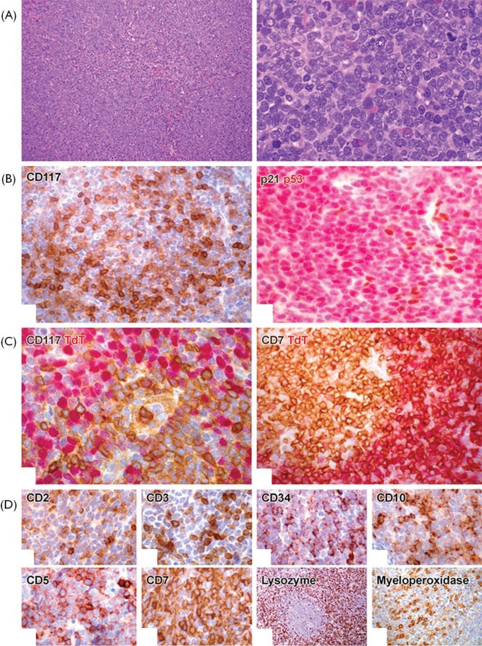
Figure 1.
Histologic features and phenotype. (A) Microscopic appearance of the lymph node biopsy shows a diffuse infiltrate of blast cells. (B,C) Immunophenotype demonstrates nodular aggregates of CD117+ terminal deoxynucleotidyl transferase (TdT)–negative myeloid blasts (left panels), surrounded by sheets of T lymphoblasts that express TdT but not CD117, although co-expression of CD7 is seen in both blast populations (right panels). (B) Double immunostaining shows expression of p53 but not p21, in keeping with mutated TP53 (right panel). (D) In addition, the T lymphoblastic lymphoma component expresses pan-T markers CD2 (weak), CD3 (weak), CD5, CD7, and precursor lymphoid markers CD34 and CD99; meanwhile, the myeloid sarcoma component expresses myeloperoxidase and CD10.

