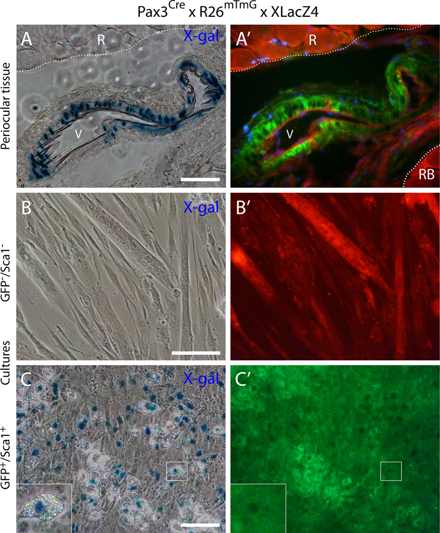
Fig. 8. Histological and cell culture analyses of periocular and EOM preparations from the double reporter mouse Pax3Cre × R26mTmG × XLacZ4.
(A) and (A’) Cross-section from periocular preparation at the level of the optic nerve, immediately posterior to the eye, depicts a blood vessel close to a rectus muscle identifying GFP+ cells at the vessel wall that co-express nuclear β-gal. R, rectus muscleRB, retractor bulbi muscleV, blood vessel. DAPI staining is shown in blue in (A’). (B-C’) Representative images of day 14 cultures of (B-B’) GFP−/Sca1− cells and (C-C’) GFP+/Sca1+ cells after staining with X-gal. Cells were isolated from EOM preparations by FACS and the specified populations were isolated from G0-G1 cells depleted of CD31+ and CD45+ cells. Insert in the lower left corner of (C) represents a higher magnification view of the region delineated by a white box in each corresponding panel, depicting an adipogenic cell expressing the XLacZ4 transgene. Scale bars in (A), 50µm, and in (B) and (C), 100µm.

