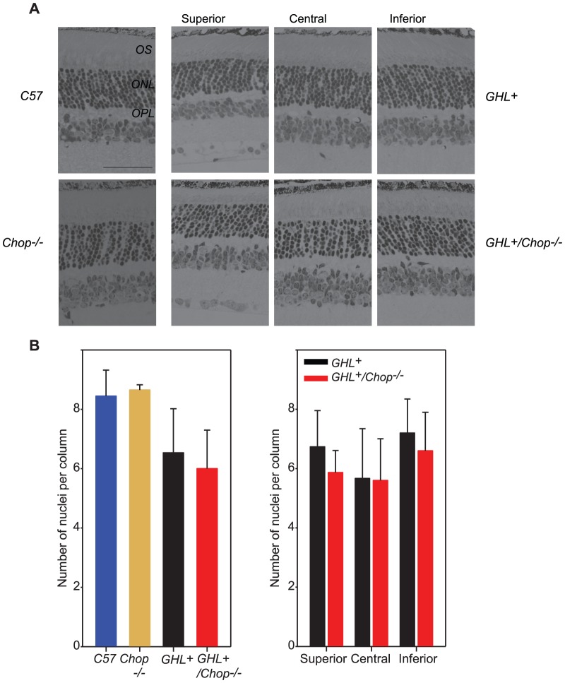
Figure 3. Histological analysis of 4 week old GHL+ and GHL+/Chop−/− retinas.
(A) Representative sections of retinas from 4 week old C57BL/6, Chop−/−, GHL+ and GHL+/Chop−/− mice (n = 2–3). (B) Mean number of photoreceptor nuclei per column counted in the ONL (left panel), and the variance in the number of photoreceptor nuclei per column counted in different zones of the retina in GHL+ and GHL+/Chop−/− mice, (right panel). Superior zone: 630 µm from the CMZ, central zone: 630 µm from optic nerve, and inferior zone: midpoint of inferior hemisphere. Error bars: ± SD OPL: outer plexiform layer, ONL: outer nuclear layer, OS: outer segment. Scale bar, 50 µm.

