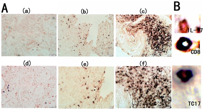
Figure 3. Expression of Tc17 cells in cervical tissues of the three groups.
(A) Immunohistochemical double staining for Tc17 cells in the control group (a and d), CIN group (b and e) and UCC group (c and f). Representative sites with low (200×, upper panels) and high (400×, lower panels) magnification were shown. (B) IL-17-producing cells were stained red (in the cytoplasm) and CD8+ cells were stained black (in the membrane). The co-expression of CD8 and IL-17 confirmed that a proportion of Tc17 cells.

