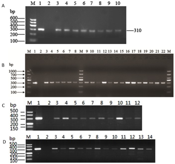
Figure 1. PCR detection of mecA-positive S. aureus isolates.
The results were analyzed by Agarose gel electrophoresis. (A) mecA-positive isolates from Gansu province. Lane M: DNA size Marker. Lane 1: positive control strain ATCC43300. Lane 2: negative control strain ATCC25923. Lanes 3–10: isolate QY4, QY6, QY8, QY10, HG2, HG3, HG4 and HG5, respectively. (B) mecA-positive isolates from Shanghai. M: DNA size Marker. Lane 1: positive control strain ATCC43300. Lane 2: negative control strain ATCC25923. Lane 3–8: isolate SX5, SX6, SX10, SX11, SX13 and SX15, respectively; and lanes 9–22: isolates SH1, SH2, SH3, SH4, SH7, SH8, SH9, SH10, SH13, SH14, SH16, SH17, SH18 and SH20, respectively. (C) mecA-positive isolates from Guizhou. M: DNA size Marker. Lane 1positive control strain ATCC43300. Lane 2: negative control strain ATCC25923. Lanes 3–12: isolates zy1, zy2, zy4, zy5, zy6, zy8, zy11, zy12, zy14, and zy15, respectively. (D). mecA-positive isolates from Sichuan. M: DNA size Marker. Lane 1positive control strain ATCC43300. Lane 2: negative control strain ATCC25923. Lanes 3–14: isolates cx1, cx2, cx5, cx6, cx8, cx9, cx10, cx13, cx14, cx17, cx18, and cx19, respectively.

