Figure 1.
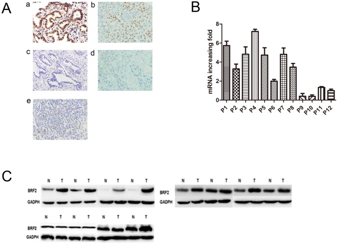
(A): The expression pattern of BRF2 in lung cancer tissues. (a) High BRF2 expression in lung adenocarcinoma. (b): High BRF2 expression in lung squamous cell carcinoma tissues. (c): Negative BRF2 expression in lung adenocarcinoma. (d): Negative BRF2 expression in lung squamous cell carcinoma tissues. (e): Intratumoral microvessels were stained as brown by the anti-CD34 monoclonal antibody in lung cancer tissues (magnification×400). (B): Quantitative real-time PCR analyses of BRF2 mRNA in twelve pairs of matched NSCLC and noncancerous tissues with GAPDH as a loading control in both panels. (C): Protein levels of BRF2 expression were evaluated by western blotting from paired noncancerous tissue and NSCLC.

