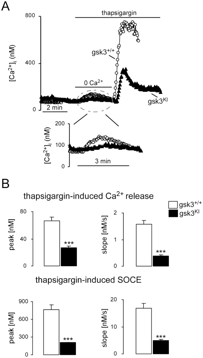
Figure 2. Thapsigargin-induced intacellular Ca2+ release and subsequent SOCE in DCs from gsk3KI and gsk3WT mice.
A. Representative original tracings showing [Ca2+]i in Fura-2/AM loaded gsk3WT (open circles) and gsk3KI (closed triangels) DCs prior to and following removal of extracellular Ca2+, addition of SERCA inhibitor thapsigargin (1 µM) and readdition of extracellular Ca2+. B. Arithmetic means ± SEM (n = 44–59) of the peak (left) and slope (right) values of [Ca2+]i increase upon Ca2+ release from intracellular stores (upper bars) and upon SOCE (lower bars) in gsk3WT DCs (white bars) and gsk3KI DCs (black bars). ***(p<0.001), unpaired t-test.

