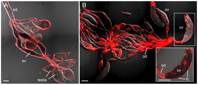
Figure 3. Accumulation of RNPs of RSV in reproductive system of viruliferous SBPHs.
At 14 days padp, reproductive organs were labelled with RSV dye RNP-rhodamine and then examined with confocal microscopy. RNP antigens were detected in the testis, seminal vesicles and ejaculatory duct of males (panel A) and in the ovarioles and oviduct of females (panel B). Inset in panel B: enlargement of boxed area to show RNPs of RSV present in the follicular cells of ovariole. Images are shown with red fluorescence (RNP antigens) under background visualized by transmitted lights. ed, ejaculatory duct; sv, seminal vesicles; fc, follicular cells of ovariole; ov, ovariole; od, oviduct. Bars, 100 µm.

