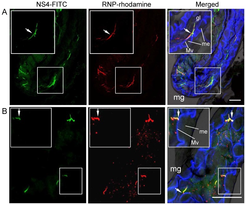
Figure 4. Association of RNPs of RSV with fibrillar inclusions of NS4 in midgut lumen of SBPH after ingestion of virus from diseased rice plants.
At 1 day padp, the alimentary canal of SBPH was labelled with NS4-FITC (green), RNP-rhodamine (red) and actin dye Phalloidin-Alexa Fluor 647 carboxylic acid (blue), and then examined with confocal microscopy. (A) Colocalization of RNPs of RSV with fibrillar inclusions of NS4 in midgut lumen. (B) Fibrillar inclusions containing complex of RNPs and NS4, attached to actin-based microvilli of epithelial cells of midgut. Arrows indicate the attachment site of fibrillar inclusion on microvilli. Insets: enlarged images of boxed areas. Images were merged with green fluorescence (NS4 antigens), red fluorescence (RNP antigens) and blue fluorescence (actin dye) under background visualized by transmitted light. mg, midgut; gl, gut lumen; Mv microvilli; me, midgut epithelium. Bars, 100 µm.

