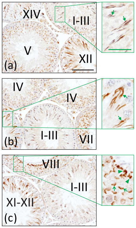
Fig. 3.
Localization of ICAM2 in the adult rat testis. A testis was embedded in paraffin, the tissue block was sectioned, and cross-sections were processed for immunohistochemistry using a monospecific ICAM2 antibody. ICAM2 localized largely to elongating and elongated spermatids [see
 in (a) and (b), insets] in all stages of the seminiferous epithelial cycle (noted by Roman numerals). Elongated spermatids in stages I–VIII were weakly immunoreactive for ICAM2. ICAM2 also localized to postacrosomal vesicles/acrosomes [see
 in (c), inset] and residual bodies [see ◀ in (c), inset]. Bar in (a), 100 μm; bar in (a), inset, 35 μm. These results are in agreement with a previously published report [212].

