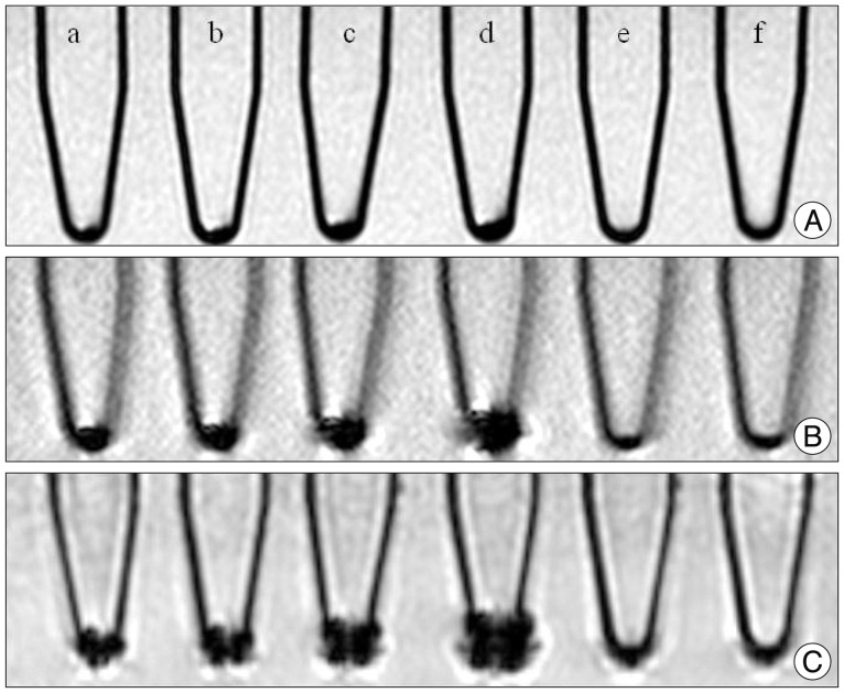
Fig. 3.
In vitro cellular MRI of PBS phantoms containing suspensions of different concentrations of SPIO labeled hBM-MSCs. A : T2 weighted image. B : T2* weighted image (GRE). C : Susceptibility weighted image. (a) 6.3×10⁴ cells/mL, (b) 1.3×10⁵ cells/mL, (c) 2.5×10⁵ cells/mL, (d) 5.0×10⁵ cells/mL, (e) unlabeled hMSC 5.0×10⁵ cells/mL, (f) no cells (normal saline). PBS : phosphate-buffered saline, hBM-MSCs : human bone marrow-derived mesenchymal stem cells, SPIO : superparamagnetic iron oxide, hMSC : human mesenchymal stem cell.

