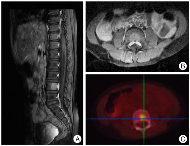
Fig. 1.

Sagittal (A) and axial (B) views of T1-weighted MRI scans with gadolinium contrast enhancement reveals a contrast-enhancing nodule of 1.6 cm in the posterior aspect of the L4 vertebral body with local FDG uptake (C) at the posterior aspect of the vertebral column, which is consistent with tumor metastasis. FDG : fluoro-deoxyglucose.
