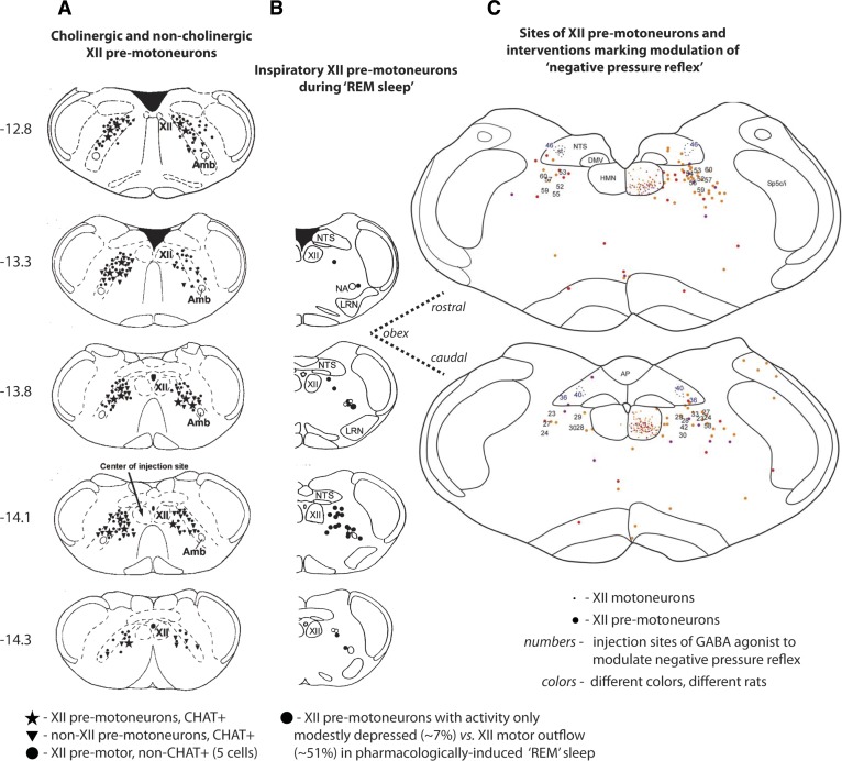
Fig. 3.
A: sections of rat brain stem showing the location of cholinergic [choline acetyltransferase positive (CHAT+)] hypoglossal (XII) premotoneurons (★) in the reticular formation lateral to the hypoglossal motor nucleus (HMN) (104). Also shown are non-XII premotor cholinergic cells (▼) and noncholinergic XII premotoneurons (●). B: the location of inspiratory XII premotoneurons that exhibit sustained or only modest depression of activity during carbachol-induced “REM sleep” [mean decrease of 7 ± 14 (SD)% from 69 ± 34 to 65 ± 37 Hz] (108). Some cells showed increased activity in the “REM” periods; in contrast, the suppression of hypoglossal motor activity (51 ± 22%) was significantly larger. C: the location of XII premotoneurons in the reticular formation adjacent to the HMN are indicated by the larger colored dots (the smaller dots indicate motoneurons in the HMN) (14). The sites of interventions with the GABAA receptor agonist muscimol to modulate the “negative pressure reflex” are also shown (sites are indicated by the numbers on the corresponding sections). Muscimol injections at and up to 500 μm rostral to the obex abolished the negative pressure reflex; injections caudal to obex did not. The sections in A and B are taken from 12.8 to 14.3 caudal to the skull reference point bregma and are plotted according to a standard brain atlas (83). The sections in C are plotted relative to obex and correspond most closely to the sections indicated by the dashed lines. AP, area postrema; DMV, dorsal motor nucleus of the vagus; NTS, nucleus of the solitary tract; LRN, lateral reticular nucleus; Amb, nucleus ambiguus. [From Volgin et al. (104), Woch et al. (108), and Chamberlin et al. (14).]

