Fig. 1.
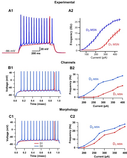
Differences in intrinsic channel properties are sufficient to describe the dichotomy in excitability between D1 and D2 medium spiny neurons (MSNs). A: experimental observations of responses of D1 and D2 MSNs to current injection. [Adapted with permission from Gertler et al. 2008.] A1: D1 MSNs have significantly higher rheobase current (median, 270 pA, n = 35) than D2 MSNs (median, 130 pA, n = 31). A2: frequency-current (F–I) plot of D1 and D2 MSNs demonstrate higher intrinsic excitability in D2 MSNs. B: models of D1 and D2 MSNs constructed with differences in intrinsic excitability reproduce electrophysiological dichotomy. The intrinsic channels with different properties are the L-type calcium channels, fast-sodium channels, slowly inactivating A-type (transient) potassium channel (KAs) and inward-rectifying potassium channels (KIR). B1: D1 and D2 MSN models have rheobase current of 275 pA and 155 pA, respectively. B2: F–I plot of these models reproduce experimentally observed differences in spiking activity. C: simulations suggest that models of D1 and D2 MSNs differentiated based solely on morphological differences do not replicate dichotomy in spiking activity. C1: D1 MSN models constructed with six primary dendrites and D2 MSN models constructed with four primary dendrites have rheobase currents of 210 pA and 160 pA, respectively. C2: the F–I plot of these MSN models does not replicate the dichotomy of D1 and D2 MSN excitability seen experimentally.

