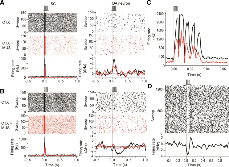
Fig. 4.
Intracollicular muscimol administration suppressed collicular and dopaminergic responses to cortical stimulation. A: raster displays (top) and PSTHs (bottom) show that collicular neurons (SC) in this animal exhibited a short-latency excitatory response to pulse trains applied to the barrel cortex (CTX; 0.1 ms, 0.6 mA; vertical dotted line). Likewise, a simultaneously recorded DA neuron showed a short-latency excitatory response to the pulse trains. After a collicular microinjection of muscimol (CTX+MUS) the collicular response to cortical stimulation was attenuated, as was the response of the DA neuron. The PSTH for the DA neuron shows the 3-point smoothed change in firing rate from baseline (ΔHz). B: as well as excitations, pulse trains applied to the barrel cortex could induce short-latency inhibitions in DA neurons. In the example shown here, intracollicular muscimol eliminated the DA neuron's response to cortical stimulation. C: trains of electrical stimuli applied to the barrel cortex produced excitatory responses in the SC to each pulse in the train (black trace). Intracollicular administration of muscimol reduced baseline activity and depressed the responses to stimulation (red trace). D: raster display (top) and PSTH (bottom) of a representative case showing that electrical stimulation of the barrel cortex with pulse trains (5 pulses at 150 Hz, 0.1 ms each, 0.6 mA) produced temporally stable responses in DA neurons.

