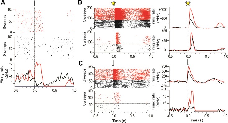
Fig. 6.
Responses of DA neurons to electrical stimulation of the barrel cortex are labile and interact with responses to visual stimulation. A: raster plots (top and middle) and a PSTH (bottom) of a DA neuron that showed a short-latency inhibitory response to single-pulse cortical stimulation of the barrel cortex (0.1 ms, 1.0 mA, 0.5 Hz) in the absence of intracollicular bicuculline (middle), which then switched to a short-latency excitatory response in the presence of bicuculline (top). The PSTH for the DA neuron shows the 3-point smoothed change in firing rate from baseline (ΔHz). B, top: raster plot and PSTH of deep layer collicular responses to whole field light flashes in the presence of intracollicular bicuculline. On trials in black flashes were preceded 2 s earlier by electrical stimulation of the barrel cortex, while on trials in red only visual stimulation was used. Bottom: raster plot and PSTH of responses in a simultaneously recorded DA neuron. In the presence of cortical stimulation, the response of the DA neuron to visual stimulation in this animal was weaker than when visual stimulation was delivered alone. C, top: raster plot and PSTH of deep-layer collicular responses to single-pulse electrical stimulation of the barrel cortex in the presence of intracollicular bicuculline. On trials in black electrical stimulation of the barrel cortex was preceded 2 s earlier by whole field light flashes, while on trials in red only electrical stimulation was used. Bottom: raster plot and PSTH of responses in a simultaneously recorded DA neuron. In the presence of visual stimulation, the response of the DA neuron to cortical stimulation in this animal was weaker than when stimulation was delivered alone.

