Figure 2.
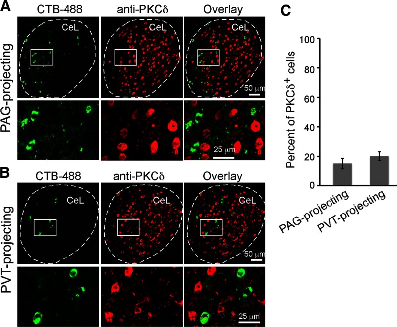
PKC-δ+ neurons do not appreciably project to PAG or PVT. A, A representative coronal brain section containing CeL, in which the PAG-projecting neurons were labeled with CTB-488 (left). PKC-δ+ neurons were recognized by an antibody (middle). Bottom, Higher-magnification images of the boxed area in the corresponding top. B, Same as A, except that the PVT-projecting CeL neurons were examined. C, Quantification of the percentage of long-range projection CeL neurons that are PKC-δ+ (n = 4 mice for each group; mean ± SEM).

