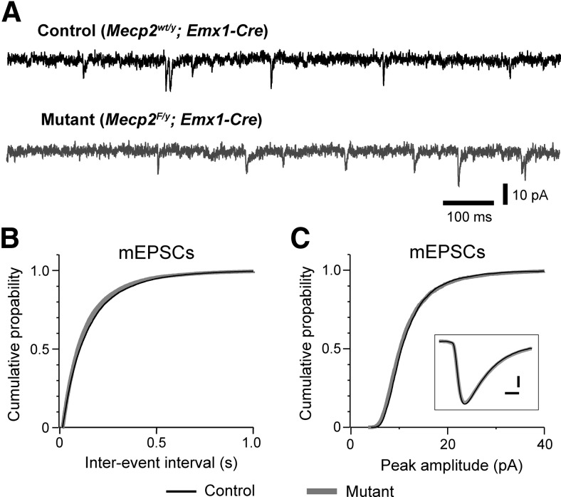
Figure 3.
Deletion of Mecp2 with Emx1-Cre had no effect on excitatory transmission in the prefrontal cortex. A, mEPSCs recorded from a control (top trace, black) and mutant (bottom trace, gray) layer 5 pyramidal neurons in mPFC at P18 in presence of TTX (0.3 μm) and picrotoxin (100 μm). B, Distributions of mEPSC intervals for mutant (black) and mutant (gray) neurons. The mean frequency was 6.3 ± 0.6 Hz for control neurons (n = 24 from 3 mice) and 6.4 ± 0.6 Hz for mutant neurons (n = 21 from 3 mice; p = 0.71). C, Distributions of mEPSC peak amplitude for control (black) and mutant (gray) neurons. The mean peak amplitude was 11.8 ± 0.4 pA for control and 12.0 ± 0.3 pA for mutant neurons (p = 0.99). Inset, Averaged mEPSCs for control (black) and mutant (gray) neurons. Scale bars, 2 pA and 2 ms. The mean decay constant of mEPSCs was 3.7 ± 0.1 ms for control and 3.8 ± 0.1 ms for mutant neurons (p = 0.89).

