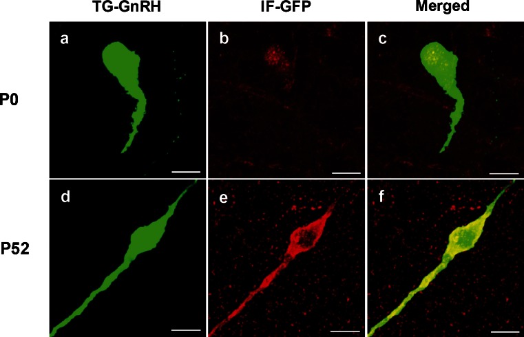
Fig. 2.
Double labeling of the green fluorescent protein immunofluroscence (IF-GFP, red) in the TG-GnRH (green) neurons in the POA of P0 (a–c) and P52 (d–f) male with the merged images (yellow). The IF-GFP staining on TG-GnRH neurons appeared weak and was localized to the nuclear region in P0 male. The IF-GFP staining was distributed across the cytoplasm and along the dendrite of the TG-GnRH neurons in P52 male. Bars 10 μm (a–f)

