
Figure 1.
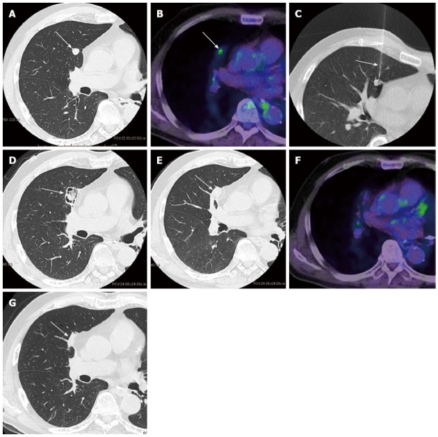
Pulmonary metastasis in a 68-year-old man with colorectal cancer treated with radiofrequency ablation. A: Computer tomography (CT) image before radiofrequency ablation (RFA) showing a tumor (arrow) 1.1 cm in size in the right middle lobe; B: Positron emission tomography (PET) image before RFA showing increased fluorodeoxyglucose (FDG) uptake by the tumor (arrow); C: CT fluoroscopic image obtained during RFA showing the treatment of the tumor with a multitined expandable electrode (arrow); D: CT image 1 mo after RFA showing cavity formation around the ablated tumor (arrow); E: CT image 3 mo after RFA showing cavity collapse and an increase in the size of the ablation zone (arrow) beyond the tumor size before RFA; F: PET image 6 mo after RFA showing the disappearance of FDG uptake; G: CT image 24 mo after RFA showing the shrinkage of the ablation zone (arrow) and its appearance as a focal atelectasis.