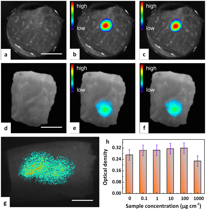
Figure 5. Anti-Stokes fluorescence tissues imaging of pork tissue with incoherent excitation.
(a–c and g) show the application of ex situ optical charging, (d–f) represent X-ray in situ charging: (a) Pre-injection autofluorescence image. (b) 60 min post-injection fluorescence image and (c) representative reproduction of (b) after 1 on/off cycling. A 980 nm laser diode was employed as the excitation source and the monitoring wavelength was set at ~700 nm. (d) Post-injection autofluorescence image without charging. (e) Post-injection fluorescence image after external X-ray charging and (f) representative reproduction of (e) after 1 on/off cycling. (g) Deep and large-area fluorescence imaging of pork tissue for an injection depth of 1 cm. A 940 nm LED was employed as the excitation source for imaging and the monitoring wavelength was set at ~700 nm. Scale bars are 15 mm for panels a–g. (h) In-vitro viability of BMSCs (bone mesenchymal stem cells) incubated with particulate Zn3Ga2Ge2O10:0.5Cr3+ as anti-Stokes probe at different concentrations for 3 days. Each data point represents the mean value of at least three independent experiments.

