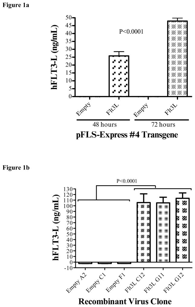
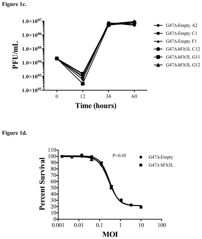
Figure 1.
Construction of G47Δ-Flt3L. a) 293T cells were transfected with the shuttle vector containing the insert with the sequence for Flt3L. ELISA assay of the supernatants demonstrates high levels of Flt3-L at 48 hours, increased further at 72 hours. Supernatant at all time points from empty plasmid-transfected 293T cells did not contain detectable Flt3L. b) Quantification of Flt3-L expression by virus-infected Vero cells. Infection of Vero cells with plaque-purified G47Δ-Flt3L resulted in elaboration of high levels of Flt3-L into the supernatant. No Flt3L was detected after infection with G47Δ without transgene expression. c) Single burst assay for viral replication. Viral replication appears to be unaffected by expression of Flt3-L. D) MTS colorimetric assay examining in vitro cytotoxicity of oncolytic virus clones. CT-2A glioma cells were infected at varying MOI’s with G47Δ-empty and G47Δ-Flt3L and assessed for viability 72 hours later. Viral expression of Flt3L did not affect the number of viable cells.


