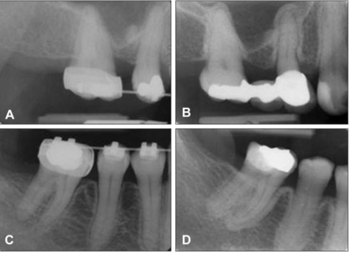
Figure 1.
Periapical radiographs of a woman, showing generalized osseous sclerosis of uniform thickness involving the cortical plate and lamina dura. A and C radiographs shows the pre-bisphosphonate therapy condition of the cortical plates and the lamina dura. B and D radiographs shows thickening of the cortical plate and lamina dura after 2 years bisphosphonate therapy. No clinical signs and symptoms were established at this initial stage

