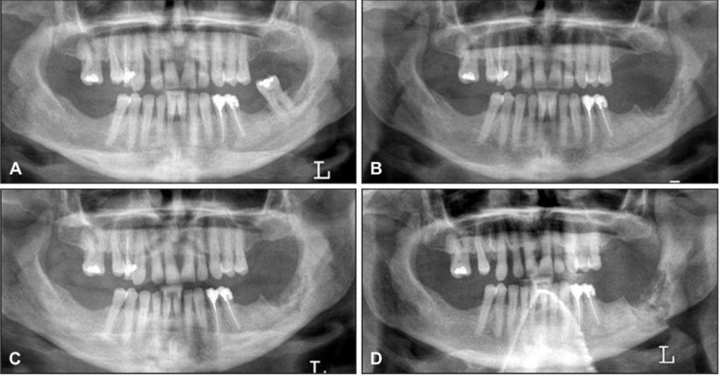
Figure 2.
A = Panoramic radiograph of a woman with breast cancer treated with zoledronate who presented with a painful tooth # 37.
B = Panoramic radiograph after 7 months, showing non healing extraction site in the left posterior mandible, absence of bone remodeling and sclerotic bone changes of the body of the mandible.
C = Panoramic radiograph after 9 months demonstrates a nonhealing extraction site in the left posterior mandible with progressive sclerosis of the left body and angle of the mandible with encroachment on the left mandibular canal.
D = Panoramic radiograph after 19 months with intervening curettage, demonstrates progression of sclerosis to pathologic fracture of the mandible.

