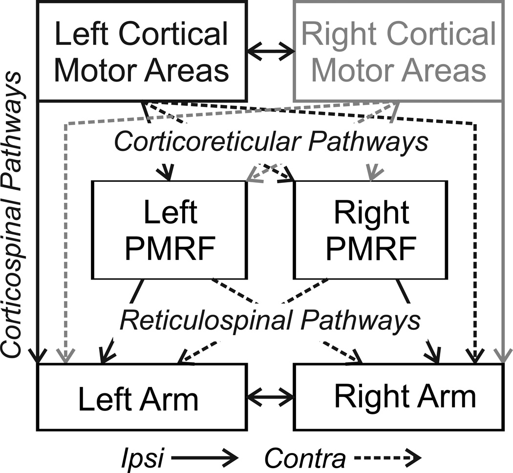
Figure 1.
Diagram of connections between cortical motor areas, the pontomedullary reticular formation (PMRF), and spinal cord segments for control of the upper limbs. Solid lines represent ipsilateral pathways, dashed lines contralateral. In the present study, stimulation was performed in the left cortical motor areas and in left and right sides of the PMRF. The grey connections show comparable projections from the right cortex.

