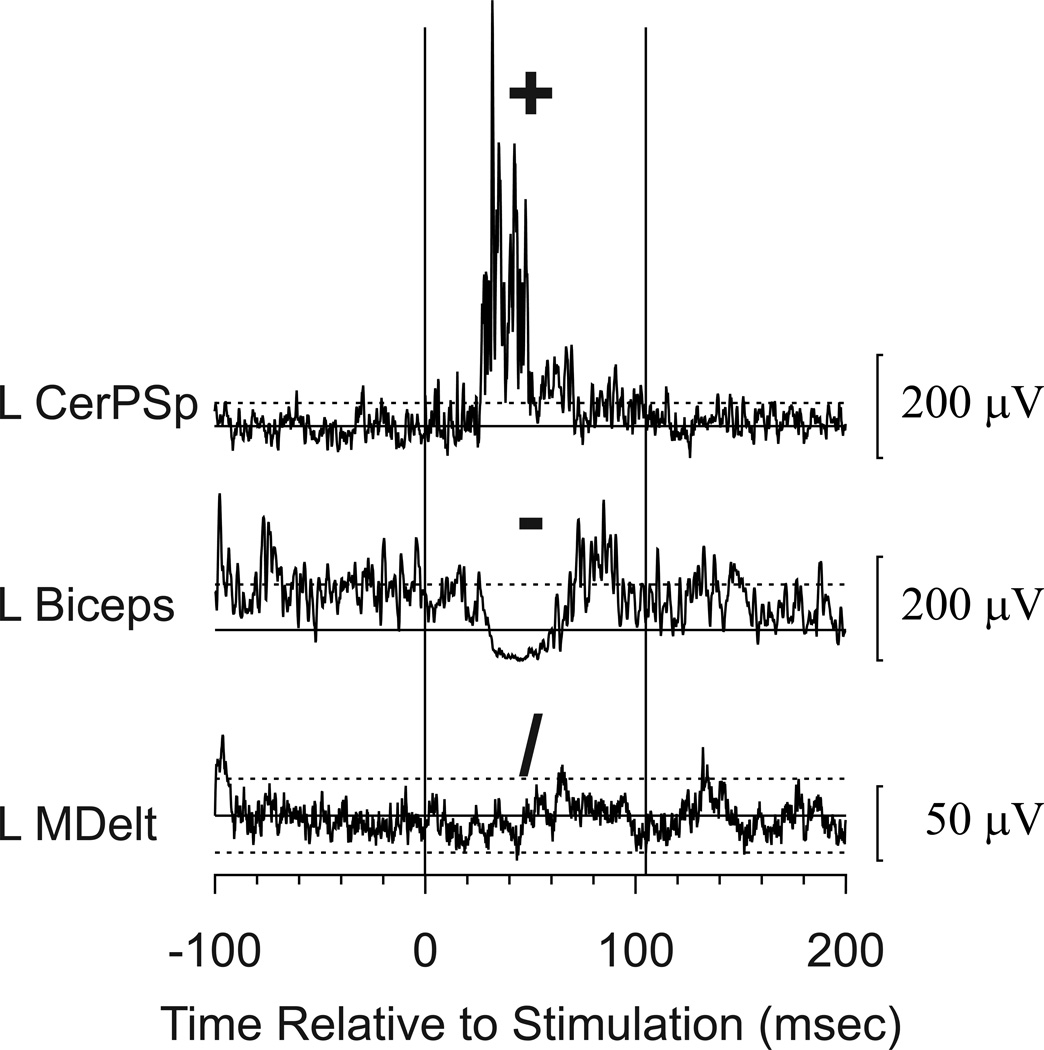
Figure 2.
Sample responses to stimulation and symbols used to code the responses. These responses were recorded during combined stimulation to the cortex and the PMRF and represent the mean EMG response to 10 stimulus trains. The solid horizontal line indicates the mean during the control period, and dashed lines show ± 2 standard deviations above or below the mean. Facilitation (coded +) was noted in the left cervical paraspinal muscle (L CerPSp), suppression (−) was noted in left Biceps (L Biceps), and there was no response (/) in the left middle deltoid (L MDelt). Several other muscles also responded to stimulation at this site, but only these examples are shown for this illustration. Calibration bars show response amplitudes.

