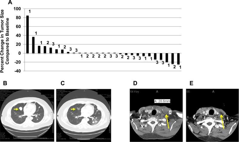
Figure 1. Clinical observations.
(A) Waterfall plot showing percent change in the sum of the diameters of tumor target lesions above or below baseline using RECIST criteria. The numbers on the graph indicate the cohort in which the patients were enrolled. Patients in cohort 1 received one cycle of 0.1mg/kg, cohort 2 0.4mg/kg and cohort 3 2mg/kg of anti-OX40. Patients 10, 18 and 23 did not have follow-up scans due to clinical progression and are PD. (B-C) Regression of a pulmonary nodule in a patient (Cohort 1) with metastatic melanoma, and progression of other nodules. Panel B, imaging before anti-OX40. Panel C, 5 months after anti-OX40 administration. (D-E) Shrinkage of a lymph node in a melanoma patient (cohort 3). Imaging in panel E was obtained 28 days after anti-OX40.

