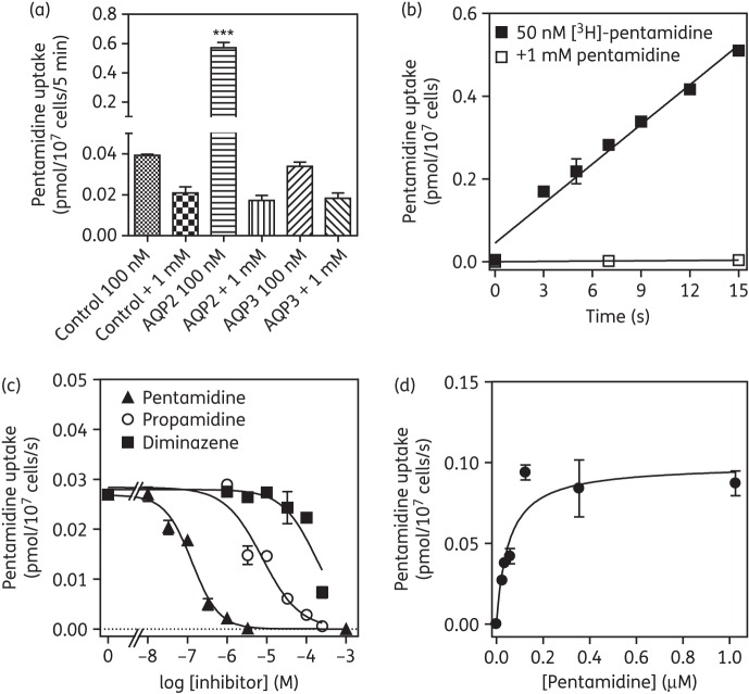
Figure 7.
Expression of T. b. brucei aquaporins in promastigotes of Leishmania mexicana. (a) Specific uptake of 100 nM [3H]pentamidine over 5 min in L. mexicana promastigotes transfected with empty pNUS vector (control), or promastigotes transfected with TbAQP2 or with TbAQP3. In each case, mediated uptake of 100 nM radiolabel was compared with total association of [3H]pentamidine with the cell pellet in the presence of a saturating concentration (1 mM) of unlabelled pentamidine. The data shown are the mean of triplicates ± SEM. ***P < 0.001 by one-way ANOVA, compared with all other datasets. (b) Time course of 50 nM [3H]pentamidine uptake, over 15 s, using L. mexicana promastigotes transformed with TbAQP2 in the presence and absence of 1 mM unlabelled pentamidine. Uptake at 50 nM pentamidine was linear (r2 = 0.98) and rapid (0.032 ± 0.002 pmol/107 cells/s, compared with 0.00026 ± 1.8 × 10⁻⁶ pmol/107 cells/s in the presence of 1 mM pentamidine). (c) Characterization of 20 nM [3H]pentamidine uptake in L. mexicana promastigotes expressing TbAQP2, in the presence of varying concentrations of unlabelled inhibitor. (d) Michaelis–Menten plot of 20 nM [3H]pentamidine uptake; conversion of pentamidine inhibition plot in (c).

