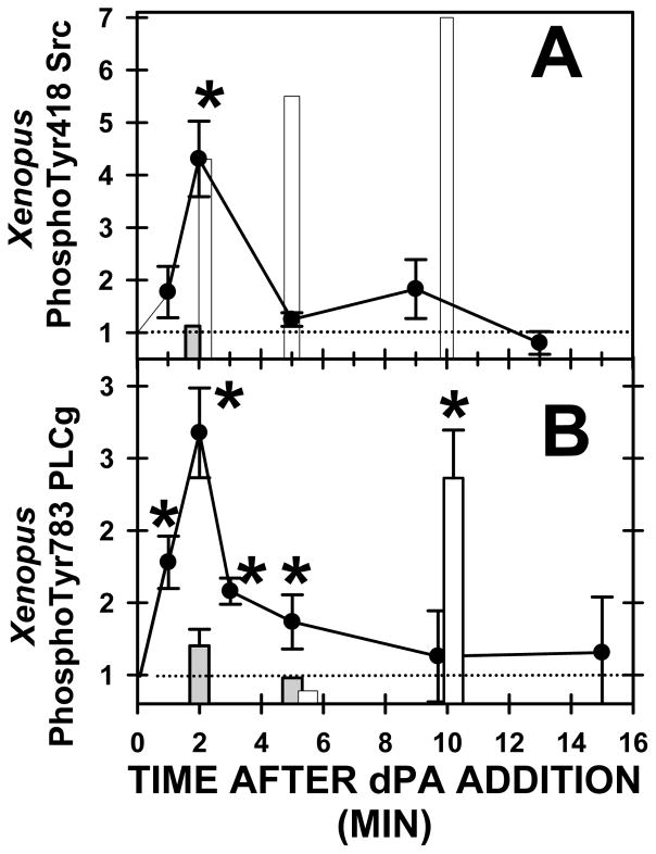
Fig. 8. Addition of exogenous PA stimulates Xenopus Src and PLCγ.
dPA (400 μM)(closed circles), control anionic lipid dPS (400 μM) (closed bars), or H2O2 (10 mM)(open bars) was added to Xenopus oocytes, phosphospecific Western blot analysis conducted and results stated relative to the control band density. (A) Xenopus phosphoSrc increased at two minutes after dPA addition, whereas dPS had no effect, and H202 stimulated Src on a much slower time course (asterisk denotes P < 0.03). Each lane has 3–9 determinations. (B) Xenopus phosphoPLCγ increased at two minutes after dPA addition, or by 10 minutes after H202 addition, however dPS addition was ineffective (asterisks denote P < 0.05).

