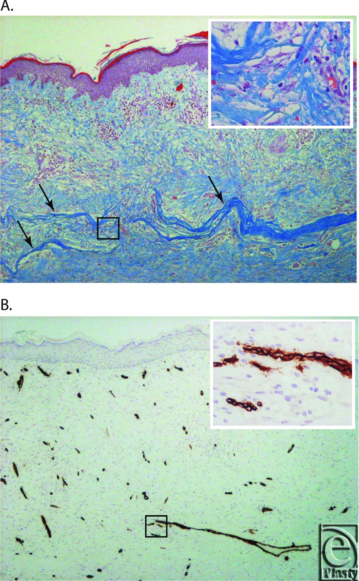
Figure 3.

(a) Gomori's Trichome stain of the excised graft from B004, 4 weeks postgraft (4× magnification). Arrows indicate the intact fragments of OFM. Insert shows a 40× magnification of the area indicated by the black square. (b) CD34 immunohistochemistry of the excised graft from B004, 4 weeks postgraft (4× magnification). Insert shows a 40× magnification of the area indicated by the black square.
