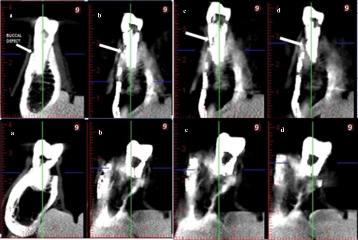
Figure 2.
Cross-sectional cone beam CT (CBCT) images of a tooth with and without buccal marginal defect. (a) Normal, without artefact reduction (AR), (b) low-mode AR, (c) medium-mode AR and (d) high-mode AR. Upper row: cross-sectional CBCT images of a tooth with buccal marginal defect. Arrows show simulated buccal defect. Lower row: cross-sectional CBCT images of a tooth without buccal marginal defect

