
Figure 1.
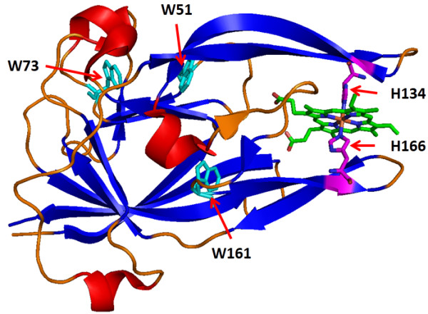
Structure of heme-bound HmuY (PDB code: 3H8T). Stick representation of tryptophan side chains with a defined color for each structure is shown. α-helices are marked in red, β-strands in blue, loops in orange, tryptophan residues in cyan, and axial histidine residues in purple.