2B.

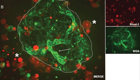
Confocal real time microscopy of a human islet surrounded by stressed single cells in suspension after islet isolation procedure. The cell surfaces are stained with WGA.While endothelial cells (see arrow) show a strong staining of the cell surface after addition of WGA, islet cells (dotted circle) are only weakly stained. Note the strong positivity for cell permeant acetoxymethylester (Rhod-2) in the stressed single cells and the high cell vitality documented by the nearly absent staining for Rhod-2 positive cells in the islet.
