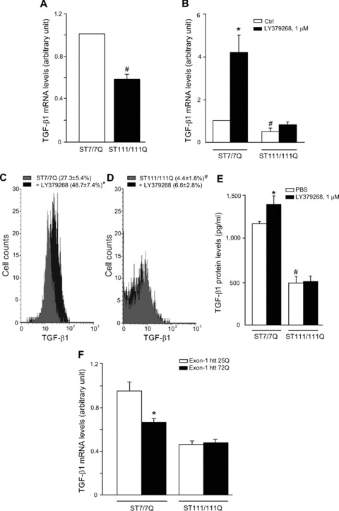
Fig 8.

Analysis of TGF-β1 in striatal-derived knock-in cells expressing full-length huntingtin with an expanded Q repeat. Real Time RT-PCR of TGF-β1 mRNA analysis (A, B) in ST7/7Q, wild-type cells or full-length mutated huntingtin ST111/111Q, mhtt cells, under basal conditions and after treatment with LY379268, 1 μM for 10 min. FACS analysis of intracellular TGF-β1 protein (C) and ELISA analysis of extracellular TGF-β1 protein wild-type and mhtt cells, under basal conditions and after treatment with LY379268, 1 μM for 10 min (D). The mean fluorescence intensity (MFI) of TGF-β1 in ST7/7Q cells (MFI: 18.1 ± 4.3, 18.0 ± 5.6, respectively) or in ST111/111Q cells (MFI: 27.3 ± 7.3, 18.0 ± 8.0, respectively) treated with PBS or LY379268 was unchanged (E). Values are means ± S.E.M. of five to six determinations. P < 0.05 versus the respective values obtained in wild-type cells treated with LY379268 (*) or versus the respective values obtained in ST7/7Q cells (#) (one-way anova+ Fisher’s PLSD). Real Time RT-PCR of TGF-β1 mRNA analysis (F) in ST7/7Q and ST111/111Q knock-in cells transfected with the unexpanded exon-1 htt 25Q or the expanded exon-1 htt 72Q. Values are means ± S.E.M. of five to six determinations. P < 0.05 versus the respective values obtained in ST7/7Q cells transfected with exon-1 htt 25Q (one-way anova+ Fisher’s PLSD).
