
Fig 5.
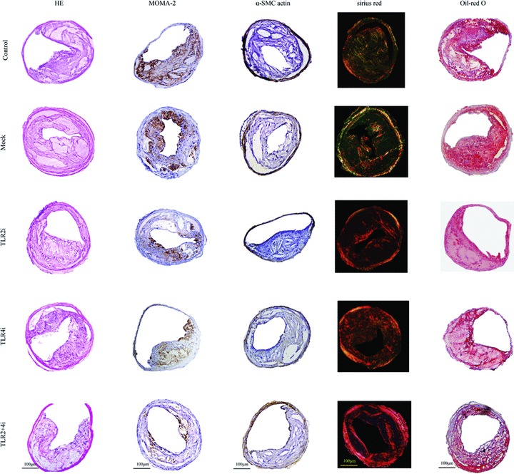
Histopathological staining showing plaque composition in the control, mock and three gene interference subgroups of mice. Cross-sections of carotid arteries in different groups were stained for haematoxylin and eosin, macrophages (MOMA-2) and vascular SMCs (α-actin), collagen (sirius red) and lipids (Oil-red O). Scale bar = 100 μm.