
Figure 3.
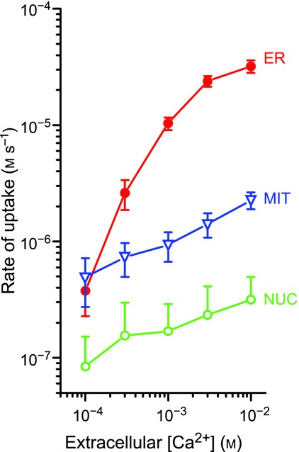
Coupling of Ca2+ entry through SOCs to calcium uptake by different organelles in HEK293 cells Measurements were performed using aequorins targeted to ER, mitochondria (MIT) and nucleus (NUC). SOC was activated by Ca2+ emptying of the ER by treatment with 10 μm tert-butyl hydroquinone (TBH), and Ca2+ entry was started by washing TBH and adding different extracellular Ca2+ concentrations (0.1 to 10 mm), as shown. The rate of entry should increase linearly with [Ca2+] (abscissa). Reproduced with permission from Alonso et al. (1998), Mol Cell Endocrinol 353, 37–44.