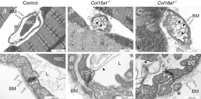
Figure 3.

Capillary morphology and endothelial cell–cell junctions in the masseter muscle Representative transmission electron micrographs of capillaries of control animals (A and D), Col15a1−/− (B and E) and Col18a1−/− mice (C and F). TEM micrographs of two mice per genotype were evaluated. Arrows point to endothelial basement membrane, white arrowheads to endothelial cell–cell junctions and black arrowheads to endothelial cell residues. Star indicates a degenerated endothelial cell. EC = endothelial cell; BM = basement membrane; P = pericyte; L = capillary lumen.
